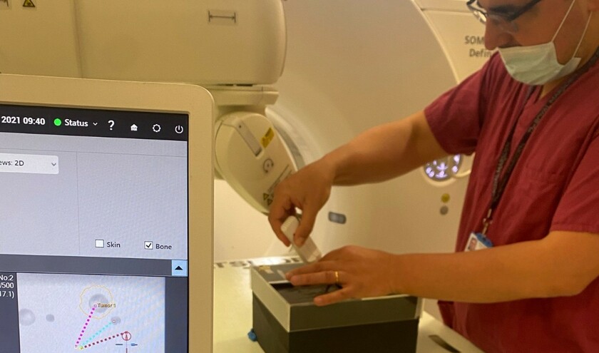
As part of the study, funded by The Royal College of Radiologists and The Royal Marsden Cancer Charity, patients with suspected retroperitoneal and pelvic sarcomas (RPS) - a rare type of soft tissue tumour - underwent an MRI a week or less before the biopsies. This was so that images of the tumour could be compared to the analysis from the biopsies.
Three ‘target regions' in one tumour, with differing imaging characteristics, were then sampled for analysis. By sampling three distinct regions, clinicians saw a more comprehensive and accurate representation of tumour biology compared to conventional single-site biopsy.
By using this technique, it was hoped that researchers would be able to better understand differences across cancer cells in different parts of the sarcoma, predict growth and gather more vital information about a particular tumour before any treatment is offered to a patient.
Dr Edward Johnston, academic consultant in oncological interventional radiology and chief investigator of the study, said: ‘This is the first time that researchers in the UK have attempted to understand the complexities of multiple tumour sites as seen on MRI before they are surgically removed.
‘Not all parts of a singular tumour behave in the same way and knowing this before treating a patient will help us to make clearer, more informed decisions about patient care.'
With further work, it is hoped that this information will lead to a very detailed understanding of how tumours appear on imaging. In the future, this information could be fed into an AI algorithm to classify tumours and forgo the need for tissue biopsy in many patients, with AI instead being able to predict tumour growth.


